
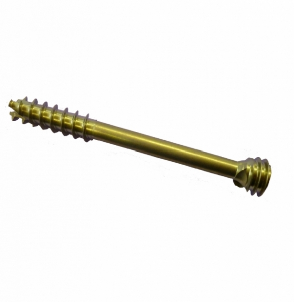
EAGLE Ø 7 screw
Product description
EAGLE Ø 7 screws are manufactured at the facilities of SARL CHOC, rue Louis Lépine, Albasud 3, 82000 Montauban, France.
EAGLE Ø 7 screws are manufactured from ISO 5832-3 standard TA6V ELI titanium. All materials are obtained from ISO 9001 certified suppliers, who provide for each titanium delivery a material conformity certificate with a physicochemical analysis.
The screws are produced in accordance with our manufacturing procedures: a manufacturing order is issued by the Production Manager. For each manufacturing order, the Production Manager assigns a four digit batch number. The manufacturing order details all the operations to be carried out on the products. At each stage, various dimensional verifications are carried out by the manufacturing operators and by the Production Manager. All inspection forms are kept with the manufacturing order.
Dimensional characteristics
EAGLE Ø 7 screws have a 7mm diameter and are available in 3 lengths of distal screw threads: 16mm (30 to 90mm long in 5mm increments), 22mm (55 to 90mm long in 5mm increments) and 28mm (70 to 90mm long in 5mm increments).
EAGLE Ø 7 screws are mainly used in the following cases:
- Calcaneal osteotomy (significant medial or lateral movement)
- Subtalar arthrodesis
- Talocrural arthrodesis
Calcaneal osteotomy
Patient in lateral decubitus, ankle to be operated on top + pneumatic tourniquet
Horizontal posterior incision.
Dissection and protection of the sural nerve.
Transverse calcaneal osteotomy guided by X-ray.
Calcaneum displacement (medial, lateral or vertical - up or down, depending on the case).
Insertion of an EAGLE Ø 7.0 mm screw kit pin, guided by X-ray from back to front between the tuberosity and the calcaneum body, maintaining reduction.
The intra bone displacement of the pin is measured with the tool provided.
A hole is then drilled through the bone over a distance of 2/3 of the length of the pin, taking care to go through the osteotomy zone using X-ray guidance.
Introduce the step drill up to the stop to prepare the screw head zone.
Finally introduce the EAGLE Ø 7.0 mm screw corresponding to the pin length measure minus 15 mm.
The EAGLE Ø 7.0 mm screw is introduced with x-ray guidance to monitor progress, so that the 2 screw threads sit either side of the osteotomy for correct stabilisation and compression.
The pin can then be removed.
X-ray guidance is then used to verify assembly stability as well as compression.
Additional immobilisation is provided by a 90° removable boot.
Talocrural arthrodesis
Patient in dorsal decubitus + pneumatic tourniquet
Anterior and medial incision on the inner side of the tibialis anterior muscle.
Opening of the articulation with protection of the neurovascular bundle, exposure of the arthritic talocrural articulation.
Execution of an extended arthrolysis expose the articular surfaces
Cartilage reduction of the articular surfaces is carried out using straight and curved chisels.
The two medial and lateral grooves are released using a reciprocating saw, removing the osteophytes.
A corresponding shortening osteotomy of the fibula is then carried out corresponding to the combined resection zone of the tibia and talus articular surfaces.
These osteotomies ensure that the articular elements fit together seamlessly.
The arthrodesis is held securely in place at the right place with two anterior compression staples.
A talus compensation anterior posterior screw is added and introduced through the anterior cortex of the tibia, obliquely downwards and to the rear.
To do this, an EAGLE Ø 7.0 mm screw pin kit is introduced by x-ray guidance according to the predefined axis taking care not to enter the subtalar joint.
The intra bone displacement of the pin is measured with the tool provided.
A hole is then drilled through the bone over a distance of 2/3 of the length of the pin, taking care to correctly transverse the joint to be treated using X-ray guidance.
Introduce the step drill up to the stop to prepare the screw head zone.
Finally introduce the EAGLE Ø 7.0 mm screw corresponding to the pin length measure minus 15 mm.
The EAGLE Ø 7.0 mm screw is introduced with x-ray guidance to monitor progress, so that the 2 screw threads are on either side of the joint that is being fused and ensure that its surfaces are properly compressed.
The pin can then be removed.
X-ray guidance is then used to verify assembly stability as well as compression.
Graft in the residual joint space that may be left.
Additional immobilisation is provided by a 90° removable boot.
Subtalar arthrodesis
Patient in lateral decubitus, ankle to be operated on top + pneumatic tourniquet
A horizontal lateral incision is made in front of the fibular malleolus end exposing the subtalar articulation.
After sectioning the inter-bone ligament, the subtalus is disassembled and the cartilaginous articular surfaces are ablated using a chisel and gouge-forceps on the posterior and anterior subtalar joint.
Preparing the congruence of surfaces and positioning them so they join edge to edge using peroperative X-ray guidance.
Insertion of the pins (provided in the EAGLE Ø 7.0 mm kit) through the sole under the heel by a short skin edge approach.
These pins are placed, under X-ray guidance, parallel one to the other in an anterior posterior position, in an upwards and frontwards direction. This allows, during the screwing process, a harmonious compression of the 2 surfaces.
The intra bone distance to be covered by the pins is then measured using the tool provided in the EAGLE Ø 7.0 mm kit (measured by subtraction).
A hole is then drilled through the bone over a distance of 2/3 of the length of the first posterior pin, taking care to pierce the joint to be treated using X-ray guidance.
Introduce the step drill up to the stop to prepare the screw head zone.
Finally introduce the EAGLE Ø 7.0 mm posterior screw corresponding to the pin length measure minus 15 mm.
The EAGLE Ø 7.0 mm screw is introduced with x-ray guidance to monitor progress, so that the 2 screw threads are on either side of the joint that is being fused and ensure that its surfaces are properly compressed.
The proximal pin can then be removed.
The same procedure is then carried out on the distal pin for the insertion of the second EAGLE Ø 7.0 mm screw.
X-ray guidance is then used to verify assembly stability as well as compression.
Graft in the residual joint space that may be left.
Additional immobilisation is provided by a 90° removable boot.
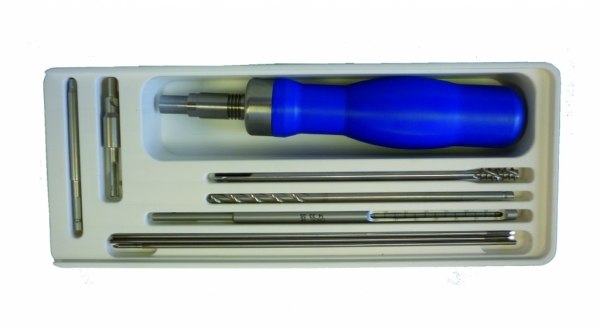
Eagle Ø 7mm screw ancillary box
14.10.97 EAGLE Ø 7 STORAGE BOX
14.32.04 EAGLE Ø 7 SCREWDRIVER HANDLE
14.30.18 EAGLE Ø 7 SCREWDRIVER SHAFT
14.37.36 EAGLE Ø 7 DRILL
14.37.35 EAGLE Ø 7 DRILL WITH STOP
14.33.34 EAGLE Ø 7 MEASURE TOOL
14.33.35 EAGLE Ø 7 TAP
14.63.45 Ø 2.5 LG 200 1 TROCAR AWL PIN
To ensure traceability, the screws are individually packaged in double blister packs, the first containing the screw and a sterilisation indicator label; the second blister contains the first blister and five traceability stickers with the screw reference, description and batch number.
The packed screws are then sent to ISOTRON France (MIN des Arnavaux- 13014 MARSEILLE) for sterilisation by gamma radiation according to the ISO 11137-02, ISO 11737-1, ISO 11737-2, EN 552, EN 556 standards and dose mapping. After sterilisation, ISOTRON returns the batch of sterilised screws with a certificate of sterilisation by gamma radiation. Once returned after sterilisation, the screws in the packaging are labelled.
The label includes the description, the reference, the production batch number, the method of sterilisation, the expiry date, the "single use" and "read instructions before use" symbols, the manufacturer's name and address and the CE 1014 markings.
The screws are packed in boxes identically labelled as above, stretch-wrapped and stored in closed cupboards, away from direct light.
Storage conditions: no special conditions.